Moving Towards Precision and Translational Medicine
Organoids, combined with biomaterial advances, are not just a scientific curiosity—they are paving the way for major developments in precision and translational medicine. By creating better, more reliable models of human tissues, scientists can use organoids to test drugs more accurately, potentially reducing the need for animal testing and improving the success rate of clinical trials. This is especially relevant when it comes to diseases that are hard to model with traditional approaches, such as certain cancers or neurodegenerative diseases(Frontiers). Looking further, biomaterial-enhanced organoids also hold the potential to personalize medicine. Imagine a future where we could take a sample of a patient’s cells, grow organoids that mimic their unique biology, and test different drugs or therapies directly on those models. This could allow for highly tailored treatment plans that offer better outcomes for patients. While we’re not quite there yet, the foundation is being laid with current research, including the use of microfluidics and advanced biomaterial scaffolding to improve nutrient flow and cellular organization in organoids(Frontiers).
A Future of Collaborative Research
The field of organoids is growing fast, and it’s an interdisciplinary effort. Advances in biomaterials are making organoids more functional, but there’s also a need for collaboration with other technologies like CRISPR gene editing, AI modeling, and proteomics. As researchers build on these technologies, organoids will become an even more powerful tool for understanding human biology and improving healthcare outcomes(Frontiers)(Frontiers).
In conclusion, biomaterials are proving to be a game-changer in the development of organoids. By mimicking the body’s natural extracellular matrix, researchers can better control how stem cells grow and organize, leading to more functional and reliable models. This opens up new opportunities in drug development, disease modeling, and personalized medicine. While challenges remain, the future of biomaterial-enhanced organoids is bright, and with continued research, we’re likely to see even greater breakthroughs in the coming years(Frontiers)(Frontiers).
- Unleashing the power of biomaterials to enhance organoid differentiation and function (Nature Methods). This article discusses how biomaterials can be used to improve the development and functionality of organoids, addressing current limitations such as variability and lack of vascularization.
- Research Progress, Challenges, and Breakthroughs of Organoids as Disease Models (Frontiers in Cell and Developmental Biology). This review outlines the advantages and disadvantages of organoids in research, including the challenges related to vascularization and the use of biomaterials(Frontiers).
- Engineering Organoid Vascularization (Biomaterials Journal). This article delves into the issues surrounding vascularization in organoids and how biomaterials and microfluidic systems can be used to solve these problems(Frontiers).
- Biomaterial stiffness regulates epithelial compartmentalization in intestinal organoids (Nature Biomedical Engineering). This study demonstrates how biomaterial stiffness influences the organization of cells in intestinal organoids(SpringerLink).
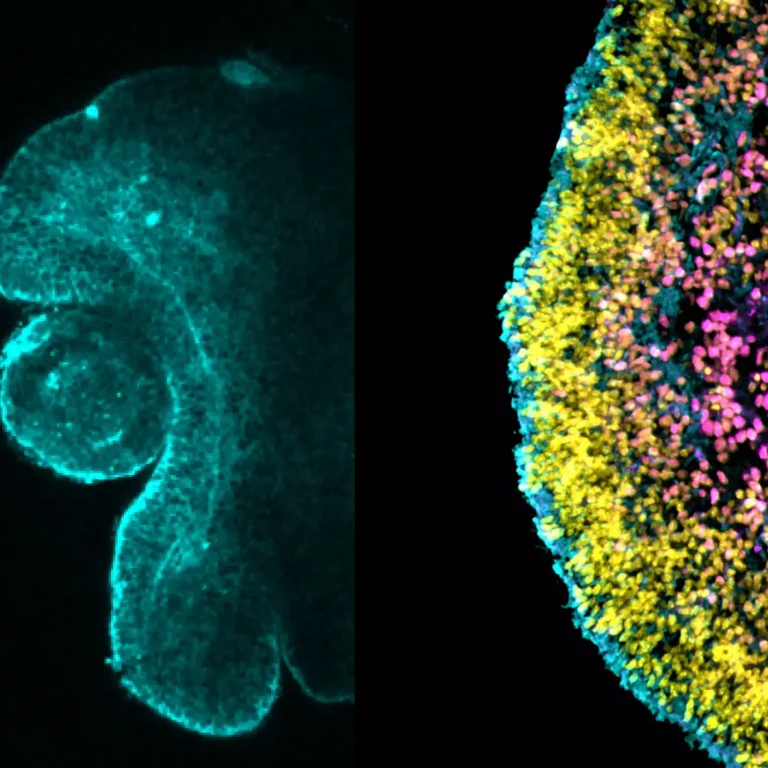
Fluorescence image of retina organoid. The retina organoid develops a typical tissue morphology (left: day 8 organoid; cyan actin-gap) and cell types which are organised into distinct layers (right: day 24 organoid; cyan rx-gfp, yellow DAPI, magenta pax6-AF594); Friedhelm Serwane
Wir benötigen Ihre Zustimmung zum Laden der Übersetzungen
Wir nutzen einen Drittanbieter-Service, um den Inhalt der Website zu übersetzen, der möglicherweise Daten über Ihre Aktivitäten sammelt. Bitte überprüfen Sie die Details in der Datenschutzerklärung und akzeptieren Sie den Dienst, um die Übersetzungen zu sehen.